- Home
- About

- Animal Owners
- Membership
- Services
- Contact us

< Back
34th Annual Scientific Meeting proceedings
Stream: LA
|
Session: In Depth: Ruminant surgery
Date/Time: 08-07-2023 (15:50 - 16:10) | Location: Theatre Hall
Date/Time: 08-07-2023 (15:50 - 16:10) | Location: Theatre Hall
Bone Surgery in Cattle
Nuss K*, Haessig M, Zimmermann I
Vetsuisse Faculty University of Zurich, Zurich, Switzerland.
Bone surgery, or surgery of the skeletal system, consists of several entities. Amongst them are trauma surgery, surgery of the joints (septic arthritis, or less commonly, luxations), surgery of the foot (which is presented elsewhere in the Ruminant section), surgical treatment of bone sequestration or osteomyelitis, and fracture treatment (Desrochers et al., 2017).
Internal fixation of long bone fractures in ruminants is applied in valuable breeding animals or pet ruminants (Nuss, 2014), and therefore still quite common in specialized clinics (Fig. 1 a-d).
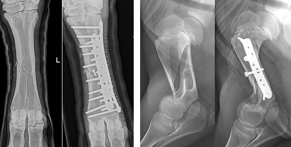
Fig1, a and b: Metacarpal fracture b and c) humerus fracture, internal fixation in a 600-kg cow and a 70 kg calf (F. Theiss, K. Nuss et al. 2023).
New technologies improve the diagnosis and therapy of bone lesions in Ruminants. For the examination of the musculoskeletal system, ultrasonography has become a valuable tool in teaching hospitals and increasingly in daily veterinary practice (Kofler et al., 2021). Beside the diagnosis of fractures and fissures (Nuss et al., 2019), sequestrum formation and bone infection are often be detected earlier with ultrasonography compared to radiography (Fig. 2 a and b).
Current CT scans offer another excellent tool for surgical planning (Fig. 2 c), because cattle can be scanned, without general anaesthesia, in only a few seconds.
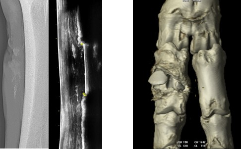
Fig. 2 a and b) sequestrum visibility with c) 2 sequestrae of P1, identified with
Radiography and Ultrasonography CT
Mandibular Fractures in Cattle
In this presentation however, the focus will be put on mandibular fractures. Mandibular fractures are among the most common fractures in cattle, as we documented in a recent study (Zimmermann et al., 2022). Medical records of cattle that were referred to the University of Zurich Veterinary Hospital because of a mandibular fracture were analysed retrospectively (Fig. 3).
In 108 cattle evaluated, the types of treatment, the short-term outcomes, the complications, and long-term outcomes were documented. Cattle that were still alive at the time of retrospective analysis (n = 11) underwent clinical and radiographic follow-up examinations.
Case load
A fall was the single most common cause of a mandibular fracture (48.1%), and a third of all cattle had a concomitant disease at the time of referral. Seventy-five cattle (49.4 %) had a single fracture, 26 had two fractures and seven had three fractures of the mandible. The molar part of the mandibular body was most commonly (40.7%) fractured followed by the diastema (23.6%), the pars incisiva (13.4%), the ramus (12.1%) and the symphysis (10.2%) of the mandible. The majority of cattle (84/108, 77.8 %) had open fractures. Forty-five cattle were euthanized or slaughtered because they had multiple fractures, or for economic reasons.

Fig 3 Numbers and distribution of mandibular fractures in 108 cattle (Zimmermann et al., 2022)
Treatment
Of 77 fractures in 63 (58.3 % of 108) treated cattle, 28 were treated with plate osteosynthesis, 25 with an external fixator, 8 with cerclage wire, and 7 using mixed techniques. Four animals were treated with fragment excision only, 4 underwent conservative treatment and one a mucosal suture (Locher et al., 2021). Complications occurred in 34 (54.0%) treated cattle; 22 had delayed fracture healing, 18 of which developed localized osteomyelitis accompanied by sequestrum formation in 14. Fifty-six of the treated cattle (88.9 %) were discharged.
The mean postoperative productive life of 45 cattle that had left their herds by the time of analysis was 46 ± 29.2 months. Thirteen of the cattle with a sequestrum remained in the herd for 15 to 92 months (mean, 47 months) and one for 2 months. The life expectancy after treatment did not differ significantly from that of the Brown Swiss and Swiss Holstein dairy cattle populations, where the cattle of this study mainly came from. Eleven cattle were available for long-term follow-up; all had a good general health status but nine had dental abnormalities including missing teeth, steps, or enamel points, all of which did not noticeably affect the body condition of the animals.
Conclusion
Surgical treatment of selected mandibular fractures had a favourable prognosis (52/63 healed, 82.5 %) in cattle.
References
- Desrochers, A., A. Steiner, D. E. Anderson, C. Guard, S. Nichols, N. G. Ducharme, K. Nuss, P.-Y. Mulon, S. Kraus, and J. A. Hill. 2017. Chapter 15 - Surgery of the Bovine Musculoskeletal System. In: Farm Animal Surgery. Fubini S. L., Ducharme N.G., 2nd ed. W.B. Saunders, pp. 344-438
- Kofler, J., A. Steiner, A. Starke, and K. Nuss. 2021. Ultrasonographic imaging of bone lesions. In: Ultrasonography of the Bovine Musculoskeletal System. Kofler J., ed. Schlütersche Verlagsanstalt, Hannover, pp.175-192.
- Locher, I., K. Nuss, D. Rediger, T. Schmid, D. Devaux, A. Steiner, and E. Marchionatti. 2021. Surgical debridement and primary closure of the oral mucosa for repair of open mandibular pars incisiva fractures in three neonatal calves. J Am Vet Med Assoc 258(11):1254-1258.
- Nuss, K. 2014. Plates, pins, and interlocking nails. Vet Clin North Am Food Anim Pract 30(1):91-126.
- Nuss, K., J. Kofler, A. Fiedler, and A. Steiner. 2019. Spezielle Diagnostik und Therapie. In: Erkrankungen der Klauen und Zehen des Rindes, eds. Fiedler A., Maierl J, and Nuss K, Thieme, Stuttgart, 2nd ed., pp. 102-174.
- Zimmermann, I., M. Haessig, and K. Nuss. 2022. [Mandibular fractures in cattle - a review of 108 cases]. Schweiz Arch Tierheilkd 164(9):609-622.
This website uses cookies to improve your experience and for traffic analysis. If you continue, we’ll assume that you agree to the use of cookies as stated in our Cookie Policy.