- Home
- About

- Animal Owners
- Membership
- Services
- Contact us

< Back
34th Annual Scientific Meeting proceedings
Stream: SA
|
Session: Neurological Injuries Associated with Pelvic Fractures
Date/Time: 05-07-2025 (09:00 - 09:30) | Location: Darwin Hall
Date/Time: 05-07-2025 (09:00 - 09:30) | Location: Darwin Hall
Decision making in Vertebral Fracture Luxation
Knell SC*
Clinic for Small Animal Surgery University of Zurich, Zurich, Switzerland.
Spinal fractures and luxations are common injuries in feline and canine trauma patients. Based on the limited available literature, approximately 16% (179/1,283) to 26% (26/100) of cats presented after a traumatic event are diagnosed with vertebral injuries. In contrast, Simpson et al. reported a lower incidence in dogs, with vertebral fractures identified in 10% (24/235) of trauma cases. An additional 12% (28/235) of dogs sustained sacral fractures or luxations, which were excluded from the primary analysis. Frequently involved is the thoracic and the lumbar area, depending on the size of the animal and the species. Large are more frequently affected in the thoracic region, whereas small dogs more often feature the lumbar region, similar to cats. Small dogs more often than cats and large dogs are affected in the cranial cervical region. In cats also the sacral or sacro-coccigeal region is injured.
Neurological assessment is crucial, as prognosis is poor in the absence of deep pain perception. Conversely, intact deep pain nociception is associated with a favorable prognosis. However, other factors must also be considered, such as neurological grade, concurrent injuries, and body weight—particularly in large dogs, where early mobilization is critical but more challenging.
At our hospital, trauma patients are predominantly cats, likely due to their greater representation in the pet population and more frequent unsupervised outdoor access, which increases the risk of injuries such as falls from height or road traffic accidents. In cats, spinal injuries frequently involve a combination of vertebral fractures and luxations, representing approximately 65% (23/49) of cases, with the lumbar spine most commonly affected. Nearly half of affected cats (12/30) are euthanized within 24 hours post-injury due to severe neurological impairment, especially when the lumbar region is involved.
Evidence guiding treatment decisions between surgical and non-surgical options remains limited, particularly in feline patients. Additionally, optimal surgical stabilization techniques are not well defined, with current practices often guided by surgeon preference and anecdotal experience rather than comparative studies.
The three-compartment model, adapted from human medicine to veterinary (primarily canine) neurosurgery, divides the vertebra into ventral, middle, and dorsal compartments. It suggests that if two or more compartments are compromised, spinal stabilization is recommended due to presumed mechanical instability.
A specific and noteworthy injury observed in dogs is the rupture of the annulus fibrosus of the intervertebral disc, without accompanying bony instability. Despite the lack of gross vertebral displacement, our clinical experience indicates significant instability, with spinal stabilization failing in 3 out of 5 cases. MRI typically reveals annular disruption, indicating a loss of disc-mediated stability, although spinal displacement is often minimal with no visible step on sagittal imaging. Therefore, diagnosis can be challenging and instability easily underestimated.
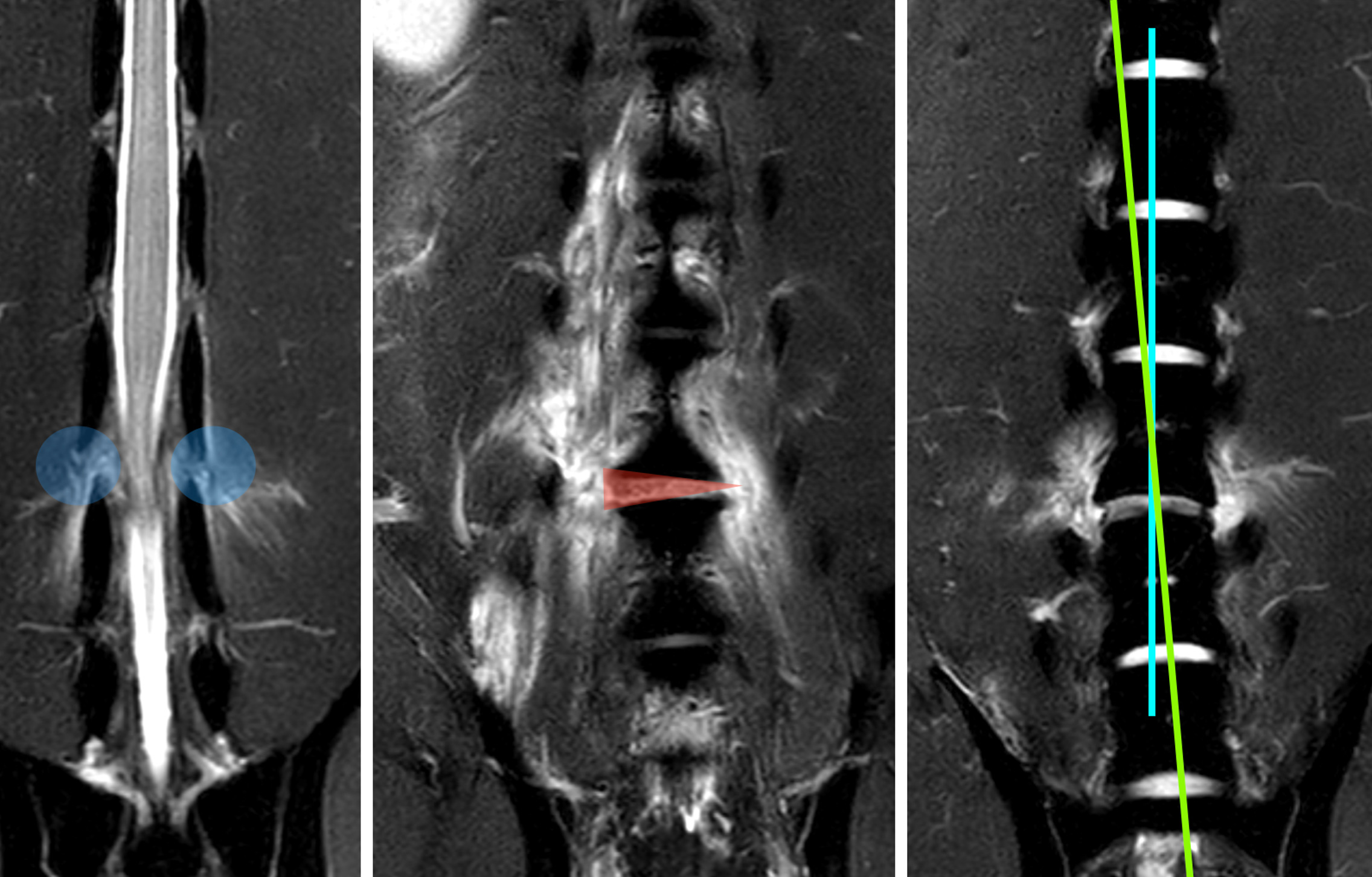
Image of a MRI study of a so called annulus rupture. Note the discrete malalignment of the spine.
Ventral stabilization techniques are the most commonly used methods for spinal fixation, typically involving plates and screws or PMMA with pins or screws. The choice between these is generally based on individual surgeon preference rather than robust comparative evidence. Biomechanically, both approaches appear to offer similar stability.
Dorsal stabilization techniques are also employed and include tension band stabilization (TS), Lubra plating, and laminar plating. These methods utilize the spinous processes or dorsal lamina for implant fixation. Dorsal techniques are favored by some surgeons due to their technical simplicity, reduced surgical time, and lower cost, despite the lack of clinical trials directly comparing them with ventral methods. TS, in particular, provides a less invasive approach and may reduce the risk of spinal canal penetration by screws or Kirschner wires (K-wires). However, the small size of feline spinous processes may limit bone purchase and predispose to implant failure.
Although TS may not fully counteract all mechanical forces acting on the spine, dorsal stabilization can offer biomechanical benefits, as demonstrated in canine models—especially for laminar stabilization—as shown by Knell et al. However, dorsal techniques are less suitable in dogs exceeding 15 kg, as failure rates increase with body weight.
This website uses cookies to improve your experience and for traffic analysis. If you continue, we’ll assume that you agree to the use of cookies as stated in our Cookie Policy.